Proposal for new catalytic roles for two invariant residues in Escherichia coli ribonuclease HI.
Kashiwagi, T., Jeanteur, D., Haruki, M., Katayanagi, K., Kanaya, S., Morikawa, K.(1996) Protein Eng 9: 857-867
- PubMed: 8931125
- DOI: https://doi.org/10.1093/protein/9.10.857
- Primary Citation of Related Structures:
1KVA, 1KVB, 1KVC - PubMed Abstract:
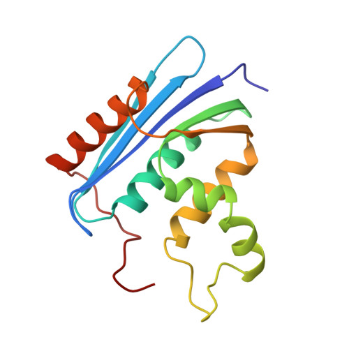
Three mutants of Escherichia coli ribonuclease HI, in which an invariant acidic residue Asp134 was replaced, were crystallized, and their three-dimensional structures were determined by X-ray crystallography. The D134A mutant is completely inactive, whereas the other two mutants, D134H and D134N, retain 59 and 90% activities relative to the wild-type, respectively. The overall structures of these three mutant proteins are identical with that of the wild-type enzyme, except for local conformational changes of the flexible loops. The ribonuclease H family has a common active site, which is composed of four invariant acidic residues (Asp10, Glu48, Asp70 and Asp134 in E.coli ribonuclease HI), and their relative positions in the mutants, even including the side-chain atoms, are almost the same as those in the wild-type. The positions of the delta-polar atoms at residue 134 in the wild-type, as well as D134H and D134N, coincide well with each other. They are located near the imidazole side chain of His124, which is assumed to participate in the catalytic reaction, in addition to the four invariant acidic residues. Combined with the pH profiles of the enzymatic activities of the two other mutants, H124A and H124A/D134N, the crystallographic results allow us to propose a new catalytic mechanism of ribonuclease H, which includes the roles for Asp134 and His124.
Organizational Affiliation:
Protein Engineering Research Institute, Osaka, Japan.