Stabilization of a strained protein loop conformation through protein engineering.
Hodel, A., Kautz, R.A., Fox, R.O.(1995) Protein Sci 4: 484-495
- PubMed: 7795531
- DOI: https://doi.org/10.1002/pro.5560040315
- Primary Citation of Related Structures:
1KDA, 1KDB, 1KDC - PubMed Abstract:
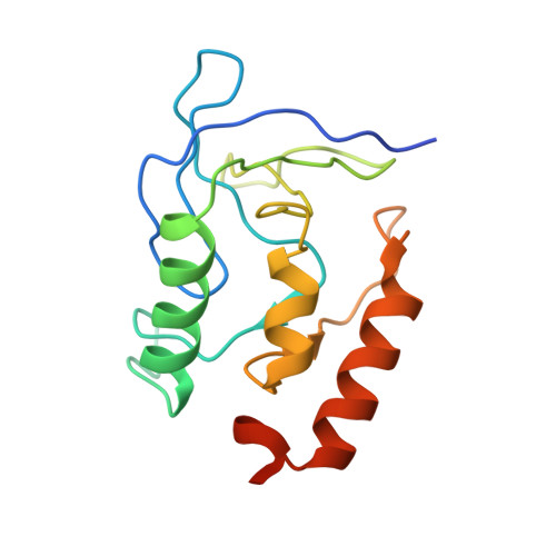
Staphylococcal nuclease is found in two folded conformations that differ in the isomerization of the Lys 116-Pro 117 peptide bond, resulting in two different conformations of the residue 112-117 loop. The cis form is favored over the trans with an occupancy of 90%. Previous mutagenesis studies have shown that when Lys 116 is replaced by glycine, a trans conformation is stabilized relative to the cis conformation by the release of steric strain in the trans form. However, when Lys 116 is replaced with alanine, the resulting variant protein is identical to the wild-type protein in its structure and in the dominance of the cis configuration. The results of these studies suggested that any nuclease variant with a non-glycine residue at position 116 should also favor the cis form because of steric requirements of the beta-carbon at this position. In this report, we present a structural analysis of four nuclease variants with substitutions at position 116. Two variants, K116E and K116M, follow the "beta-carbon" hypothesis by favoring the cis form. Furthermore, the crystal structure of K116E is nearly identical to that of the wild-type protein. Two additional variants, K116D and K116N, provide exceptions to this simple "beta-carbon" rule in that the trans conformation is stabilized relative to the cis configuration by these substitutions. Crystallographic data indicate that this stabilization is effected through the addition of tertiary interactions between the side chain of position 116 with the surrounding protein and water structure. The detailed trans conformation of the K116D variant appears to be similar to the trans conformation observed in the K116G variant, suggesting that these two mutations stabilize the same conformation but through different mechanisms.
Organizational Affiliation:
Department of Molecular Biophysics and Biochemistry, Yale University, New Haven, Connecticut 06511, USA.