Mechanistic and structural contributions of critical surface and internal residues to cytochrome c electron transfer reactivity.
Rafferty, S.P., Guillemette, J.G., Berghuis, A.M., Smith, M., Brayer, G.D., Mauk, A.G.(1996) Biochemistry 35: 10784-10792
- PubMed: 8718869
- DOI: https://doi.org/10.1021/bi960430v
- Primary Citation of Related Structures:
1IRV, 1IRW - PubMed Abstract:
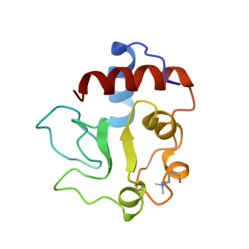
The influence of mutations in two conserved regions of yeast iso-1-cytochrome c believed to be critical to the mechanism of cytochrome c electron transfer reactions has been investigated. The variants Asn52Ala, Tyr67Phe, Ile75Met, and Thr78Gly involve perturbation of critical hydrogen-bonding interactions with an internal water molecule (Wat166) and have been studied in terms of their electrochemical properties and the kinetics with which they are reduced by Fe(EDTA)2- and oxidized by Co(phen)3(3+). In parallel studies, the Co(phen)3(3+) oxidation kinetics of Tyr, Leu, Ile, Ala, Ser, and Gly variants of the phylogenetically conserved residue Phe82 have been studied and correlated with previous electrochemical and kinetic results. To assist mechanistic interpretation of these results, the three-dimensional structures of the Asn52Ala and Ile75Met ferrocytochrome c variants have been determined. The reduction potentials of the variants modified in the region of Wat166 were at least 33 mV (pH 6, 25 degrees C, and mu = 0.1 M) lower than that of the wild-type protein. Electron transfer reactivity of this family of variants in both the oxidation and reduction reactions was increased as much as 10-fold over that of the wild-type cytochrome. On the other hand, the reactivity of the position-82 variants in both oxidation and reduction depended on the structural characteristics of the oxidation-reduction reagent with which they reacted, and this reactivity was related to the nature of the residue at this position. These findings have been interpreted as demonstrating that the principal influence of modification at position-82 arises from changes in the nature of reactant-protein interaction at the surface of the protein and in maintaining the high reduction potential of the cytochrome while the principal influence of internal modifications near Wat166 results from alteration of the reorganization energy for the oxidation state-linked conformational change defined by crystallographic analysis of the wild-type protein.
Organizational Affiliation:
Department of Biochemistry and Molecular Biology, University of British Columbia, Vancouver, Canada.