1HQ3

CRYSTAL STRUCTURE OF THE HISTONE-CORE-OCTAMER IN KCL/PHOSPHATE


Experimental Data Snapshot

  • Method: X-RAY DIFFRACTION
  • Resolution: 2.15 Å
  • R-Value Free: 0.253 
  • R-Value Work: 0.214 

wwPDB Validation   3D Report Full Report


This is version 1.3 of the entry. See complete history


Literature

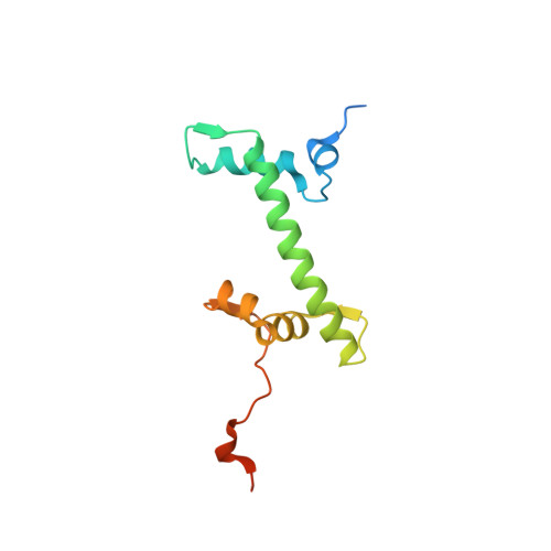
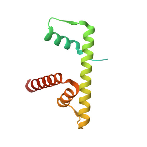
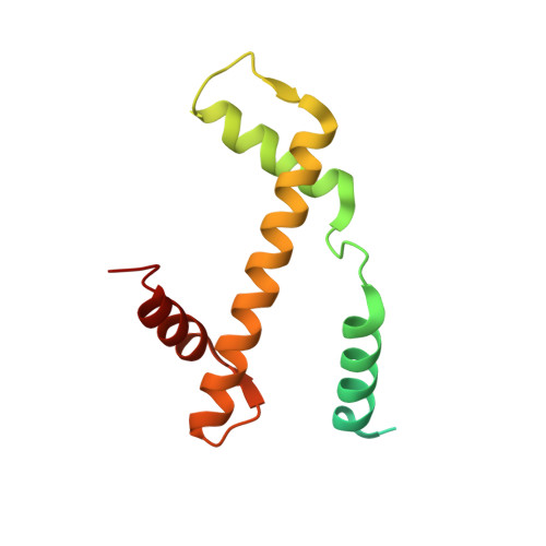
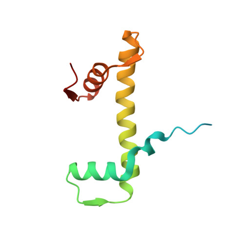
Structure of the histone-core octamer in KCl/phosphate crystals at 2.15 A resolution.

Chantalat, L.Nicholson, J.M.Lambert, S.J.Reid, A.J.Donovan, M.J.Reynolds, C.D.Wood, C.M.Baldwin, J.P.

(2003) Acta Crystallogr D Biol Crystallogr 59: 1395-1407

  • DOI: https://doi.org/10.1107/s0907444903011880
  • Primary Citation of Related Structures:  
    1HQ3

  • PubMed Abstract: 

    The structure of the native chicken histone octamer, crystallized in 2 M KCl, 1.35 M potassium phosphate pH 6.9, has been refined at 2.15 A resolution to a final R factor of 21.4% and an R(free) of 25.2%. Unique crystal-packing interactions between histone-core octamers are strong and one of them (area 4000 A(2)) involves two chloride ions and direct interactions between six acidic amino-acid residues on one octamer and the equivalent number of basic residues on the next. These interactions are on the structured part of the octamer (not involving tails). Five phosphate ions, 23 chloride ions and 437 water molecules have been identified in the structure. The phosphate and some chloride ions bind to basic amino-acid residues that interact with DNA in the nucleosome. The binding of most of the anions and the packing interactions are unique to these crystals. In other respects, and including the positions of four chloride ions, the octamer structure is very close to that of octamers in nucleosome-core particle crystals, particularly with respect to 'docking' sequences of the histone H2As and H4s. These sequences together with the H2B-H4 four-helix bundles stabilize the histone structure in the nucleosome and prevent the dissociation of the (H2A-H2B) dimers from the (H3-H4)(2) tetramer. Possible reasons why this happens at high salt in the absence of DNA are given.


  • Organizational Affiliation

    Structural Biology, Galderma RandD, 635 Route des Lucioles, BP 87F-06902 Sophia Antipolis CEDEX, France.


Macromolecules
Find similar proteins by:  (by identity cutoff)  |  3D Structure
Entity ID: 1
MoleculeChains Sequence LengthOrganismDetailsImage
HISTONE H2A-IV
A, E
129Gallus gallusMutation(s): 0 
UniProt
Find proteins for P02263 (Gallus gallus)
Explore P02263 
Go to UniProtKB:  P02263
Entity Groups  
Sequence Clusters30% Identity50% Identity70% Identity90% Identity95% Identity100% Identity
UniProt GroupP02263
Sequence Annotations
Expand
  • Reference Sequence
Find similar proteins by:  (by identity cutoff)  |  3D Structure
Entity ID: 2
MoleculeChains Sequence LengthOrganismDetailsImage
HISTONE H2B
B, F
126Gallus gallusMutation(s): 0 
UniProt
Find proteins for P0C1H5 (Gallus gallus)
Explore P0C1H5 
Go to UniProtKB:  P0C1H5
Entity Groups  
Sequence Clusters30% Identity50% Identity70% Identity90% Identity95% Identity100% Identity
UniProt GroupP0C1H5
Sequence Annotations
Expand
  • Reference Sequence
Find similar proteins by:  (by identity cutoff)  |  3D Structure
Entity ID: 3
MoleculeChains Sequence LengthOrganismDetailsImage
HISTONE H3
C, G
136Gallus gallusMutation(s): 0 
UniProt
Find proteins for P84229 (Gallus gallus)
Explore P84229 
Go to UniProtKB:  P84229
Entity Groups  
Sequence Clusters30% Identity50% Identity70% Identity90% Identity95% Identity100% Identity
UniProt GroupP84229
Sequence Annotations
Expand
  • Reference Sequence
Find similar proteins by:  (by identity cutoff)  |  3D Structure
Entity ID: 4
MoleculeChains Sequence LengthOrganismDetailsImage
HISTONE H4-VI
D, H
103Gallus gallusMutation(s): 0 
UniProt
Find proteins for P62801 (Gallus gallus)
Explore P62801 
Go to UniProtKB:  P62801
Entity Groups  
Sequence Clusters30% Identity50% Identity70% Identity90% Identity95% Identity100% Identity
UniProt GroupP62801
Sequence Annotations
Expand
  • Reference Sequence
Small Molecules
Ligands 2 Unique
IDChains Name / Formula / InChI Key2D Diagram3D Interactions
PO4
Query on PO4

Download Ideal Coordinates CCD File 
I [auth A],
O [auth B],
Q [auth C],
X [auth E],
Y [auth E]
PHOSPHATE ION
O4 P
NBIIXXVUZAFLBC-UHFFFAOYSA-K
CL
Query on CL

Download Ideal Coordinates CCD File 
AA [auth E]
BA [auth F]
CA [auth G]
DA [auth G]
EA [auth G]
AA [auth E],
BA [auth F],
CA [auth G],
DA [auth G],
EA [auth G],
FA [auth G],
GA [auth H],
HA [auth H],
IA [auth H],
J [auth A],
K [auth A],
L [auth A],
M [auth A],
N [auth A],
P [auth B],
R [auth C],
S [auth C],
T [auth C],
U [auth D],
V [auth D],
W [auth D],
Z [auth E]
CHLORIDE ION
Cl
VEXZGXHMUGYJMC-UHFFFAOYSA-M
Experimental Data & Validation

Experimental Data

  • Method: X-RAY DIFFRACTION
  • Resolution: 2.15 Å
  • R-Value Free: 0.253 
  • R-Value Work: 0.214 
  • Space Group: P 65
Unit Cell:
Length ( Å )Angle ( ˚ )
a = 158.56α = 90
b = 158.56β = 90
c = 102.48γ = 120
Software Package:
Software NamePurpose
DENZOdata reduction
SCALEPACKdata scaling
AMoREphasing
CNSrefinement

Structure Validation

View Full Validation Report


Entry History 

Deposition Data

Revision History  (Full details and data files)

  • Version 1.0: 2001-01-24
    Type: Initial release
  • Version 1.1: 2008-04-27
    Changes: Version format compliance
  • Version 1.2: 2011-07-13
    Changes: Version format compliance
  • Version 1.3: 2023-08-09
    Changes: Data collection, Database references, Derived calculations, Refinement description