1DRF

CRYSTAL STRUCTURE OF HUMAN DIHYDROFOLATE REDUCTASE COMPLEXED WITH FOLATE


Experimental Data Snapshot

  • Method: X-RAY DIFFRACTION
  • Resolution: 2.00 Å
  • R-Value Observed: 0.189 

wwPDB Validation   3D Report Full Report


This is version 1.3 of the entry. See complete history


Literature

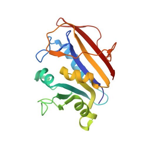
Crystal structure of human dihydrofolate reductase complexed with folate.

Oefner, C.D'Arcy, A.Winkler, F.K.

(1988) Eur J Biochem 174: 377-385

  • DOI: https://doi.org/10.1111/j.1432-1033.1988.tb14108.x
  • Primary Citation of Related Structures:  
    1DRF

  • PubMed Abstract: 

    The crystal structure of recombinant human dihydrofolate reductase with folate bound in the active site has been determined and the structural model refined at 0.2-nm resolution. Preliminary studies of the binding of the inhibitors methotrexate and trimethoprim to the human apoenzyme have been performed at 0.35-nm resolution. The conformations of the chemically very similar ligands folate and methotrexate, one a substrate the other a potent inhibitor, differ substantially in that their pteridine rings are in inverse orientations relative to their p-aminobenzoyl-L-glutamate moieties. Methotrexate binding is similar to that previously observed in two bacterial enzymes but is quite different from that observed in the enzyme from a mouse lymphoma cell line [Stammers et al. (1987) FEBS Lett. 218, 178-184]. The geometry of the polypeptide chain around the folate binding site in the human enzyme is not consistent with conclusions previously drawn with regard to the species selectivity of the inhibitor trimethoprim [Matthews et al. (1985) J. Biol. Chem. 260, 392-399].


  • Organizational Affiliation

    Central Research Units, F. Hoffmann-La Roche & Co. Ltd, Basel, Switzerland.


Macromolecules
Find similar proteins by:  (by identity cutoff)  |  3D Structure
Entity ID: 1
MoleculeChains Sequence LengthOrganismDetailsImage
DIHYDROFOLATE REDUCTASE186Homo sapiensMutation(s): 0 
EC: 1.5.1.3
UniProt & NIH Common Fund Data Resources
Find proteins for P00374 (Homo sapiens)
Explore P00374 
Go to UniProtKB:  P00374
PHAROS:  P00374
GTEx:  ENSG00000228716 
Entity Groups  
Sequence Clusters30% Identity50% Identity70% Identity90% Identity95% Identity100% Identity
UniProt GroupP00374
Sequence Annotations
Expand
  • Reference Sequence
Small Molecules
Ligands 2 Unique
IDChains Name / Formula / InChI Key2D Diagram3D Interactions
FOL
Query on FOL

Download Ideal Coordinates CCD File 
D [auth A]FOLIC ACID
C19 H19 N7 O6
OVBPIULPVIDEAO-LBPRGKRZSA-N
SO4
Query on SO4

Download Ideal Coordinates CCD File 
B [auth A],
C [auth A]
SULFATE ION
O4 S
QAOWNCQODCNURD-UHFFFAOYSA-L
Binding Affinity Annotations 
IDSourceBinding Affinity
FOL Binding MOAD:  1DRF Kd: 111 (nM) from 1 assay(s)
Experimental Data & Validation

Experimental Data

  • Method: X-RAY DIFFRACTION
  • Resolution: 2.00 Å
  • R-Value Observed: 0.189 
  • Space Group: H 3
Unit Cell:
Length ( Å )Angle ( ˚ )
a = 86.29α = 90
b = 86.29β = 90
c = 76.98γ = 120
Software Package:
Software NamePurpose
PROLSQrefinement

Structure Validation

View Full Validation Report


Entry History 

Deposition Data

Revision History  (Full details and data files)

  • Version 1.0: 1992-01-15
    Type: Initial release
  • Version 1.1: 2008-03-03
    Changes: Version format compliance
  • Version 1.2: 2011-07-13
    Changes: Version format compliance
  • Version 1.3: 2024-02-07
    Changes: Data collection, Database references, Derived calculations, Other