


1BRR
X-RAY STRUCTURE OF THE BACTERIORHODOPSIN TRIMER/LIPID COMPLEX
- PDB DOI: https://doi.org/10.2210/pdb1BRR/pdb
- Classification: PROTON TRANSPORT
- Organism(s): Halobacterium salinarum
- Mutation(s): No
- Membrane Protein: Yes PDBTMMemProtMD
- Deposited: 1998-07-28 Released: 1998-09-30
Experimental Data Snapshot
- Method: X-RAY DIFFRACTION
- Resolution: 2.90 Å
- R-Value Free: 0.299
- R-Value Work: 0.256
- R-Value Observed: 0.256

This is version 3.1 of the entry. See complete history.

Macromolecules
Find similar proteins by:
(by identity cutoff) | 3D Structure
Entity ID: 1 | |||||
|---|---|---|---|---|---|
| Molecule | Chains | Sequence Length | Organism | Details | Image |
| PROTEIN (BACTERIORHODOPSIN) | 247 | Halobacterium salinarum | Mutation(s): 0 Membrane Entity: Yes | 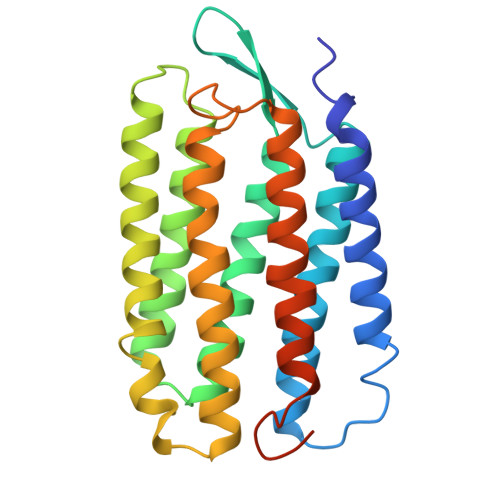 | |
UniProt | |||||
Find proteins for P02945 (Halobacterium salinarum (strain ATCC 700922 / JCM 11081 / NRC-1)) Explore P02945 Go to UniProtKB: P02945 | |||||
Entity Groups | |||||
| Sequence Clusters | 30% Identity50% Identity70% Identity90% Identity95% Identity100% Identity | ||||
| UniProt Group | P02945 | ||||
Sequence AnnotationsExpand | |||||
| |||||
Oligosaccharides

Small Molecules
| Ligands 5 Unique | |||||
|---|---|---|---|---|---|
| ID | Chains | Name / Formula / InChI Key | 2D Diagram | 3D Interactions | |
| ARC Query on ARC | G [auth A] H [auth A] I [auth A] M [auth B] N [auth B] | 3,7,11,15-TETRAMETHYL-HEXADECAN-1-OL C20 H42 O AJAKLDUGVSKVDG-UFYCRDLUSA-N |  | ||
| RET Query on RET | F [auth A], L [auth B], S [auth C] | RETINAL C20 H28 O NCYCYZXNIZJOKI-OVSJKPMPSA-N |  | ||
| BGC Query on BGC | K [auth B] | beta-D-glucopyranose C6 H12 O6 WQZGKKKJIJFFOK-VFUOTHLCSA-N |  | ||
| OCT Query on OCT | Q [auth B] | N-OCTANE C8 H18 TVMXDCGIABBOFY-UHFFFAOYSA-N |  | ||
| GOL Query on GOL | J [auth A], R [auth B], W [auth C] | GLYCEROL C3 H8 O3 PEDCQBHIVMGVHV-UHFFFAOYSA-N |  | ||
| Modified Residues 1 Unique | |||||
|---|---|---|---|---|---|
| ID | Chains | Type | Formula | 2D Diagram | Parent |
| PCA Query on PCA | A, B, C | L-PEPTIDE LINKING | C5 H7 N O3 |  | GLN |
Experimental Data & Validation
Experimental Data
- Method: X-RAY DIFFRACTION
- Resolution: 2.90 Å
- R-Value Free: 0.299
- R-Value Work: 0.256
- R-Value Observed: 0.256
- Space Group: C 1 2 1
Unit Cell:
| Length ( Å ) | Angle ( ˚ ) |
|---|---|
| a = 120.52 | α = 90 |
| b = 105.96 | β = 94.94 |
| c = 80.19 | γ = 90 |
| Software Name | Purpose |
|---|---|
| X-PLOR | model building |
| X-PLOR | refinement |
| DENZO | data reduction |
| CCP4 | data scaling |
| X-PLOR | phasing |
Entry History
Deposition Data
- Released Date: 1998-09-30 Deposition Author(s): Essen, L.-O., Siegert, R., Oesterhelt, D.
Revision History (Full details and data files)
- Version 1.0: 1998-09-30
Type: Initial release - Version 1.1: 2008-04-27
Changes: Version format compliance - Version 1.2: 2011-07-13
Changes: Non-polymer description, Version format compliance - Version 2.0: 2019-12-25
Changes: Advisory, Data collection, Database references, Derived calculations, Polymer sequence - Version 3.0: 2020-07-29
Type: Remediation
Reason: Carbohydrate remediation
Changes: Advisory, Atomic model, Data collection, Derived calculations, Structure summary - Version 3.1: 2023-08-09
Changes: Data collection, Database references, Derived calculations, Refinement description, Structure summary
