1BRG

CRYSTALLOGRAPHIC ANALYSIS OF PHE->LEU SUBSTITUTION IN THE HYDROPHOBIC CORE OF BARNASE


Experimental Data Snapshot

  • Method: X-RAY DIFFRACTION
  • Resolution: 2.20 Å
  • R-Value Work: 0.168 
  • R-Value Observed: 0.168 

wwPDB Validation   3D Report Full Report


This is version 1.6 of the entry. See complete history


Literature

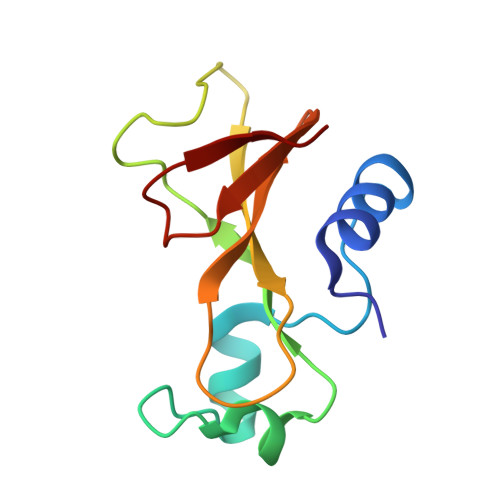
Crystallographic analysis of Phe-->Leu substitution in the hydrophobic core of barnase.

Chen, Y.W.Fersht, A.R.Henrick, K.

(1995) Acta Crystallogr D Biol Crystallogr 51: 220-231

  • DOI: https://doi.org/10.1107/S0907444994008851
  • Primary Citation of Related Structures:  
    1BRG

  • PubMed Abstract: 

    The crystal structure of a barnase mutant, Phe-->Leu7 has been determined to 2.2 A resolution. No structural rearrangement is observed near the mutated residue. The F7L mutation is highly destabilizing and this is caused by the loss of extensive van der Waals contacts that wild-type Phe7 made with its neighbouring residues, and the exposure of a large hydrophobic pocket on the surface of the protein. The side-chain conformations of the mutated Leu7 residue have torsion angles chi(1) ranging from -138 degrees to -168 degrees and chi(2) ranging from +16 degrees to +70 degrees, for the three molecules in the asymmetric unit. These angles do not agree with the most frequently observed conformations in the protein side-chain rotamer library [Ponder & Richards (1987). J. Mol. Biol. 193, 775-791]. However, when compared to a more recent 'backbone-dependent' rotamer library [Dunbrack & Karplus (1993). J. Mol. Biol. 230, 543-574], the side-chain conformation of Leu7 agrees well with that of the most frequently observed rotamers. The side-chain conformation of Leu7 was found to be dictated by two factors: it has the lowest conformational energy and it buries the most hydrophobic surface area.


  • Organizational Affiliation

    Centre for Protein Engineering, Medical Research Council Centre, Cambridge, England.


Macromolecules
Find similar proteins by:  (by identity cutoff)  |  3D Structure
Entity ID: 1
MoleculeChains Sequence LengthOrganismDetailsImage
BARNASE
A, B, C
108Bacillus amyloliquefaciensMutation(s): 0 
EC: 3.1.27
UniProt
Find proteins for P00648 (Bacillus amyloliquefaciens)
Explore P00648 
Go to UniProtKB:  P00648
Entity Groups  
Sequence Clusters30% Identity50% Identity70% Identity90% Identity95% Identity100% Identity
UniProt GroupP00648
Sequence Annotations
Expand
  • Reference Sequence
Small Molecules
Ligands 1 Unique
IDChains Name / Formula / InChI Key2D Diagram3D Interactions
ZN
Query on ZN

Download Ideal Coordinates CCD File 
D [auth C]ZINC ION
Zn
PTFCDOFLOPIGGS-UHFFFAOYSA-N
Experimental Data & Validation

Experimental Data

  • Method: X-RAY DIFFRACTION
  • Resolution: 2.20 Å
  • R-Value Work: 0.168 
  • R-Value Observed: 0.168 
  • Space Group: P 32
Unit Cell:
Length ( Å )Angle ( ˚ )
a = 58.723α = 90
b = 58.723β = 90
c = 82.026γ = 120
Software Package:
Software NamePurpose
X-PLORmodel building
PROLSQrefinement
X-PLORrefinement
X-PLORphasing

Structure Validation

View Full Validation Report


Entry History 

Deposition Data

Revision History  (Full details and data files)

  • Version 1.0: 1994-06-22
    Type: Initial release
  • Version 1.1: 2008-03-04
    Changes: Version format compliance
  • Version 1.2: 2011-07-13
    Changes: Version format compliance
  • Version 1.3: 2017-11-29
    Changes: Derived calculations, Other
  • Version 1.4: 2019-07-17
    Changes: Data collection, Refinement description
  • Version 1.5: 2019-08-14
    Changes: Data collection, Refinement description
  • Version 1.6: 2024-02-07
    Changes: Data collection, Database references, Derived calculations