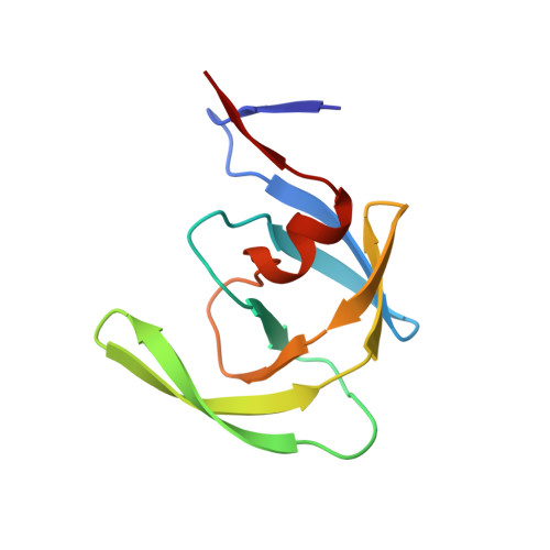
1BDL

HIV-1 (2:31-37) PROTEASE COMPLEXED WITH INHIBITOR SB203386


Experimental Data Snapshot

  • Method: X-RAY DIFFRACTION
  • Resolution: 2.80 Å
  • R-Value Free: 0.255 
  • R-Value Work: 0.201 

Starting Model: experimental
View more details

wwPDB Validation   3D Report Full Report


Ligand Structure Quality Assessment 


This is version 1.5 of the entry. See complete history


Literature

Structural role of the 30's loop in determining the ligand specificity of the human immunodeficiency virus protease.

Swairjo, M.A.Towler, E.M.Debouck, C.Abdel-Meguid, S.S.

(1998) Biochemistry 37: 10928-10936

  • DOI: https://doi.org/10.1021/bi980784h
  • Primary Citation of Related Structures:  
    1BDL, 1BDQ, 1BDR

  • PubMed Abstract: 

    The structural basis of ligand specificity in human immunodeficiency virus (HIV) protease has been investigated by determining the crystal structures of three chimeric HIV proteases complexed with SB203386, a tripeptide analogue inhibitor. The chimeras are constructed by substituting amino acid residues in the HIV type 1 (HIV-1) protease sequence with the corresponding residues from HIV type 2 (HIV-2) in the region spanning residues 31-37 and in the active site cavity. SB203386 is a potent inhibitor of HIV-1 protease (Ki = 18 nM) but has a decreased affinity for HIV-2 protease (Ki = 1280 nM). Crystallographic analysis reveals that substitution of residues 31-37 (30's loop) with those of HIV-2 protease renders the chimera similar to HIV-2 protease in both the inhibitor binding affinity and mode of binding (two inhibitor molecules per protease dimer). However, further substitution of active site residues 47 and 82 has a compensatory effect which restores the HIV-1-like inhibitor binding mode (one inhibitor molecule in the center of the protease active site) and partially restores the affinity. Comparison of the three chimeric protease structures with those of HIV-1 and SIV proteases complexed with the same inhibitor reveals structural changes in the flap regions and the 80's loops, as well as changes in the dimensions of the active site cavity. The study provides structural evidence of the role of the 30's loop in conferring inhibitor specificity in HIV proteases.


  • Organizational Affiliation

    Department of Structural Biology and Molecular Biology, SmithKline Beecham Pharmaceuticals, King of Prussia, Pennsylvania 19406, USA.


Macromolecules
Find similar proteins by:  (by identity cutoff)  |  3D Structure
Entity ID: 1
MoleculeChains Sequence LengthOrganismDetailsImage
HIV-1 PROTEASE
A, B
99Human immunodeficiency virus 1Mutation(s): 7 
Gene Names: HIV-1 PROTEASE
EC: 3.4.23.16
UniProt
Find proteins for P04587 (Human immunodeficiency virus type 1 group M subtype B (isolate BH5))
Explore P04587 
Go to UniProtKB:  P04587
Entity Groups  
Sequence Clusters30% Identity50% Identity70% Identity90% Identity95% Identity100% Identity
UniProt GroupP04587
Sequence Annotations
Expand
  • Reference Sequence
Small Molecules
Ligands 1 Unique
IDChains Name / Formula / InChI Key2D Diagram3D Interactions
IM1
Query on IM1

Download Ideal Coordinates CCD File 
C [auth A],
D [auth B]
(2R,4S,5S,1'S)-2-PHENYLMETHYL-4-HYDROXY-5-(TERT-BUTOXYCARBONYL)AMINO-6-PHENYL HEXANOYL-N-(1'-IMIDAZO-2-YL)-2'-METHYLPROPANAMIDE
C31 H42 N4 O4
QAHXABIFJPGWDD-WKAQUBQDSA-N
Binding Affinity Annotations 
IDSourceBinding Affinity
IM1 PDBBind:  1BDL Ki: 1410 (nM) from 1 assay(s)
Experimental Data & Validation

Experimental Data

  • Method: X-RAY DIFFRACTION
  • Resolution: 2.80 Å
  • R-Value Free: 0.255 
  • R-Value Work: 0.201 
  • Space Group: P 61
Unit Cell:
Length ( Å )Angle ( ˚ )
a = 63.41α = 90
b = 63.41β = 90
c = 83.55γ = 120
Software Package:
Software NamePurpose
X-PLORmodel building
X-PLORrefinement
DENZOdata reduction
SCALEPACKdata scaling
X-PLORphasing

Structure Validation

View Full Validation Report


Ligand Structure Quality Assessment 


Entry History 

Deposition Data

Revision History  (Full details and data files)

  • Version 1.0: 1998-08-12
    Type: Initial release
  • Version 1.1: 2008-03-03
    Changes: Version format compliance
  • Version 1.2: 2011-07-13
    Changes: Version format compliance
  • Version 1.3: 2021-11-03
    Changes: Database references, Derived calculations, Other
  • Version 1.4: 2023-08-02
    Changes: Refinement description
  • Version 1.5: 2024-05-22
    Changes: Data collection