1A0O

CHEY-BINDING DOMAIN OF CHEA IN COMPLEX WITH CHEY


Experimental Data Snapshot

  • Method: X-RAY DIFFRACTION
  • Resolution: 2.95 Å
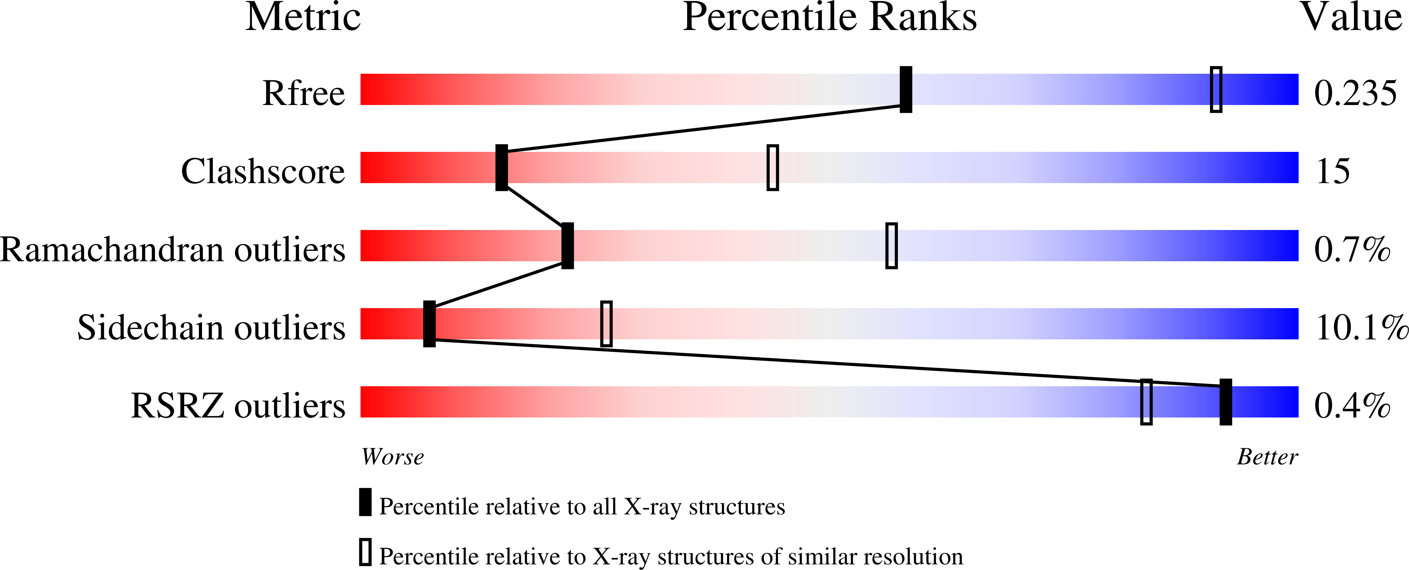
  • R-Value Free: 0.235 
  • R-Value Work: 0.187 
  • R-Value Observed: 0.187 

wwPDB Validation   3D Report Full Report


This is version 1.3 of the entry. See complete history


Literature

Structure of the CheY-binding domain of histidine kinase CheA in complex with CheY.

Welch, M.Chinardet, N.Mourey, L.Birck, C.Samama, J.P.

(1998) Nat Struct Biol 5: 25-29

  • DOI: https://doi.org/10.1038/nsb0198-25
  • Primary Citation of Related Structures:  
    1A0O

  • PubMed Abstract: 

    Bacterial adaptation to the environment is accomplished through the coordinated activation of specific sensory receptors and signal processing proteins. Among the best characterized of these pathways are those which employ the two-component paradigm. In these systems, signal transmission is mediated by Mg(2+)-dependent phospho-relay reactions between histidine auto-kinases and phospho-accepting receiver domains in response-regulator proteins. Although this mechanism of activation is common to all response-regulators, detrimental cross-talk between different two-component pathways within the same cell is minimized through the use of specific recognition domains. Here, we report the crystal structure, at 2.95 A resolution, of the response regulator of bacterial chemotaxis, CheY, bound to the recognition domain from its cognate histidine kinase, CheA. The structure suggests that molecular recognition, in this low affinity complex (KD = 2 microM), may also contribute to the mechanism of CheY activation.


  • Organizational Affiliation

    Institut de Pharmacologie et de Biologie Structurale CNRS, UPR9062, Toulouse, France.


Macromolecules
Find similar proteins by:  (by identity cutoff)  |  3D Structure
Entity ID: 1
MoleculeChains Sequence LengthOrganismDetailsImage
CHEY
A, C, E, G
128Escherichia coliMutation(s): 0 
UniProt
Find proteins for P0AE67 (Escherichia coli (strain K12))
Explore P0AE67 
Go to UniProtKB:  P0AE67
Entity Groups  
Sequence Clusters30% Identity50% Identity70% Identity90% Identity95% Identity100% Identity
UniProt GroupP0AE67
Sequence Annotations
Expand
  • Reference Sequence
Find similar proteins by:  (by identity cutoff)  |  3D Structure
Entity ID: 2
MoleculeChains Sequence LengthOrganismDetailsImage
CHEA
B, D, F, H
134Escherichia coliMutation(s): 0 
EC: 2.7.3
UniProt
Find proteins for P07363 (Escherichia coli (strain K12))
Explore P07363 
Go to UniProtKB:  P07363
Entity Groups  
Sequence Clusters30% Identity50% Identity70% Identity90% Identity95% Identity100% Identity
UniProt GroupP07363
Sequence Annotations
Expand
  • Reference Sequence
Experimental Data & Validation

Experimental Data

  • Method: X-RAY DIFFRACTION
  • Resolution: 2.95 Å
  • R-Value Free: 0.235 
  • R-Value Work: 0.187 
  • R-Value Observed: 0.187 
  • Space Group: P 1 21 1
Unit Cell:
Length ( Å )Angle ( ˚ )
a = 53.9α = 90
b = 156.97β = 91.7
c = 65.97γ = 90
Software Package:
Software NamePurpose
SHARPphasing
X-PLORmodel building
X-PLORrefinement
MOSFLMdata reduction
CCP4data scaling
SCALAdata scaling
X-PLORphasing

Structure Validation

View Full Validation Report


Entry History 

Deposition Data

Revision History  (Full details and data files)

  • Version 1.0: 1998-12-30
    Type: Initial release
  • Version 1.1: 2008-03-24
    Changes: Version format compliance
  • Version 1.2: 2011-07-13
    Changes: Version format compliance
  • Version 1.3: 2023-08-02
    Changes: Database references, Derived calculations, Refinement description